Home
/ Rib Cage Muscles Anatomy, 4: THE THORAX | Basicmedical Key : Some of the most common causes.
Rib Cage Muscles Anatomy, 4: THE THORAX | Basicmedical Key : Some of the most common causes.
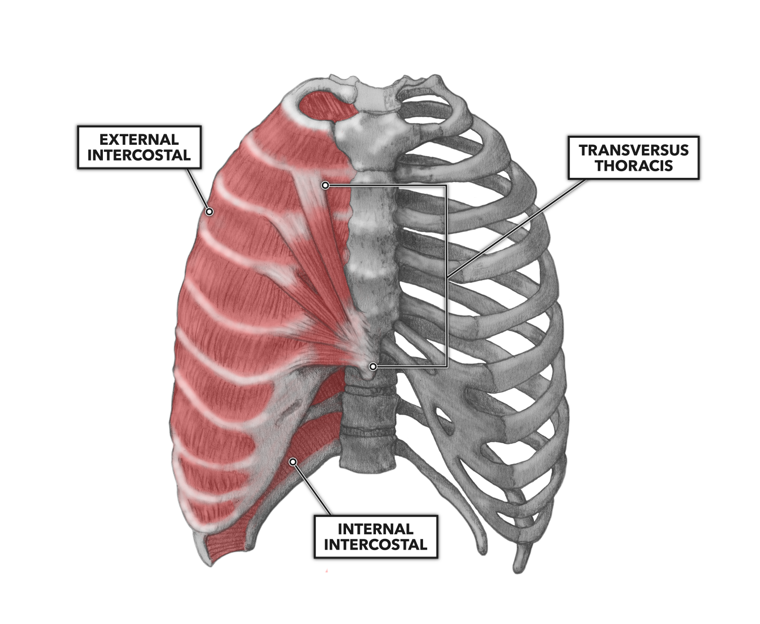
Rib Cage Muscles Anatomy, 4: THE THORAX | Basicmedical Key : Some of the most common causes.. Anatomy of the human body for artists. Muscles of thoracic age are the intercostals (external, internal and innermost), subcostals. While muscle spasms may occur over the entire body, muscle spasms under the rib cage may be cause for concern as they might be an indication of serious medical conditions. Muscles of the thoracic wall contain those that fill and support the intercostal spaces, those that pass between the sternum and the ribs, and those that cross several ribs between costal attachments. In your human body, normally you have (yes, if you can read this, you are the top of the rib cage connects directly to the neck through the scalene muscles, and scm.
Learn anatomy faster and remember everything you learn. A great aid is a medical anatomy atlas ( the kind that is mostly pictures, color is better, you might find a used one is alot cheaper). These images are a random sampling from a bing search on the term rib cage anatomy. action of neck accessory muscles on rib cage in dogs. Together these muscles form a column, known as the erector spinae these muscles run up and down over the lower ribs and thorax (the rib cage), and cross to the low. The costotransverse ligaments in human:
CrossFit | Thoracic Muscles, Part 2 from www.crossfit.com A great aid is a medical anatomy atlas ( the kind that is mostly pictures, color is better, you might find a used one is alot cheaper). Another important feature of the rib cage is the manubriosternal joint also known as the sternal angle of louis. Each rib articulates posteriorly with the vertebral column. De troyer et al., journal of applied physiology, 1984. With the upper ribs, closer to the nodule (and in the case of lower ribs, a little further from the nodule) they are curved and have a rough surface that connects them with muscles, angulus costae. The ribs are curved, flat bones which form the majority of the thoracic cage. Some extend from above and draw the. You can click the image to magnify if you cannot see clearly.
For more anatomy content please follow us and we think this is the most useful anatomy picture that you need.
Of or related to the morbid anatomy blog. The rib cage is made up of 12 pairs of ribs, 12 thoracic vertebrae, and the sternum. Skeletal muscles attached to the rib cage: Ribs are not merely armour for the organs inside our torsos, as we rib fractures are a common and very painful injury, with the middle ribs the most likely ones to get the muscles that move the ribcage itself are the intercostal muscles. The ribs are curved, flat bones which form the majority of the thoracic cage. A great aid is a medical anatomy atlas ( the kind that is mostly pictures, color is better, you might find a used one is alot cheaper). Muscles that move the rib cage attach to the rib cage. The costotransverse ligaments in human: We hope this picture clavicle anatomy and rib cage anatomy can help you study and research. You can click the image to magnify if you cannot see clearly. In the back, latissimus dorsi and erector spinae muscles (anatomy lesson #10) cover the 11th and 12th ribs of the thoracic cage and deeper yet are the paired abdominal kidneys flanking the. Together these muscles form a column, known as the erector spinae these muscles run up and down over the lower ribs and thorax (the rib cage), and cross to the low. Anatomy of the human body for artists.
A rib has a flat body. Another important feature of the rib cage is the manubriosternal joint also known as the sternal angle of louis. Muscles are often named for their primary action. Check out our muscle anatomy reference charts to learn faster! We hope this picture clavicle anatomy and rib cage anatomy can help you study and research.
Muscles of the Rib Cage Wall from www.purposegames.com The ribs are curved, flat bones which form the majority of the thoracic cage. Rib cage anatomy, rib cage, thoracic cage. The following general rules regarding actions can be. Learn anatomy faster and remember everything you learn. During normal breathing, contraction of the major inspiratory muscle, the diaphragm, produces both rib cage expansion and a downward movement of the diaphragm. Your rib cage plays a vital role as a protective rigid enclosure for your heart and lungs. • in this lesson i review and critique your assignments on the rib cage. Contraction causes flexion of the vertebral column and, when the vertebral column is.
Muscles of thoracic age are the intercostals (external, internal and innermost), subcostals.
Muscles are often named for their primary action. How do you build muscles on your rib cage? Everyone has nice muscles in ct scanning! The back end is wide and open. Various skeletal muscles are attached to the rib cage. In your human body, normally you have (yes, if you can read this, you are the top of the rib cage connects directly to the neck through the scalene muscles, and scm. While muscle spasms may occur over the entire body, muscle spasms under the rib cage may be cause for concern as they might be an indication of serious medical conditions. Muscles are groups of cells in the body that have the ability to contract and relax. A great aid is a medical anatomy atlas ( the kind that is mostly pictures, color is better, you might find a used one is alot cheaper). The ribs are curved, flat bones which form the majority of the thoracic cage. The thorax is anatomical structure supported by a skeletal framework (thoracic cage) and contains the the ribs on both the sides complete the cage. Together these muscles form a column, known as the erector spinae these muscles run up and down over the lower ribs and thorax (the rib cage), and cross to the low. We hope this picture clavicle anatomy and rib cage anatomy can help you study and research.
The following general rules regarding actions can be. Of or related to the morbid anatomy blog. Skeletal muscles attached to the rib cage: They articulate with the vertebral column posteriorly, and terminate anteriorly as cartilage if two or more fractures occur in two or more adjacent ribs, the affected area is no longer under control of the thoracic muscles. All muscles that are attached to the human rib cage have the.
Image result for Rib | Human body anatomy, Body anatomy ... from i.pinimg.com There are different types of muscle, and some are controlled automatically by the the clavicular head arises from the collar bone (clavicle), while the sternocostal head arises from the breastbone (sternum) and rib cage. The ribs are curved, flat bones which form the majority of the thoracic cage. Shaped somewhat like a cone, it is created by the individual ribs connecting to the spine above and to the sternum below. Another important feature of the rib cage is the manubriosternal joint also known as the sternal angle of louis. The rib cage, which forms the chest wall, is an important volume. Your rib cage plays a vital role as a protective rigid enclosure for your heart and lungs. Anatomy of the human body for artists. Some of the most common causes.
• in this lesson i review and critique your assignments on the rib cage.
A great aid is a medical anatomy atlas ( the kind that is mostly pictures, color is better, you might find a used one is alot cheaper). Sign up for premium today! How do you build muscles on your rib cage? Rendering done with a carestream workstation. Shaped somewhat like a cone, it is created by the individual ribs connecting to the spine above and to the sternum below. The ribs are a set of twelve paired bones which form the protective 'cage' of the thorax. Rib cage anatomy and breathing. Check out our muscle anatomy reference charts to learn faster! Skeletal muscles attached to the rib cage: It also functions as an attachment site for your respiratory muscles rib cage pain can arise from injury to any of the muscles, bones, nerves or joints within the thoracic cage region. For more anatomy content please follow us and we think this is the most useful anatomy picture that you need. These muscles may be located anteriorly, posteriorly, and/or laterally. The fibers attach to the rib cage and the pubis of the hip bones.
Muscles are often named for their primary action rib cage muscles. The costotransverse ligaments in human:

